If you want to feel infinitesimal and insignificant, you need only look at a galaxy-clogged night sky—or a small, magnified slice of brain matter. The dusty streaks of supernovas and dead stars in the observable universe are only shrunk by the slimy clump of neurons and synapses in a human skull. Is the whole of my being—my Gaussian memories, my anxiety disorders and tics, my capacity for intimacy or destruction—really self-contained within this electrified slushball of meat?
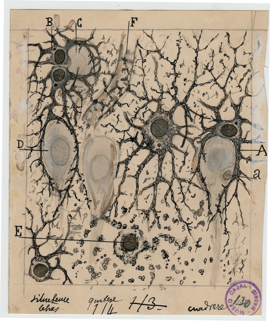
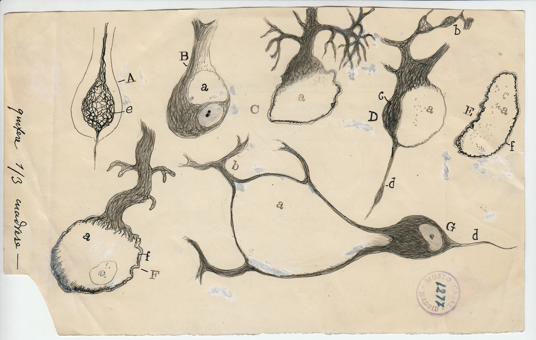
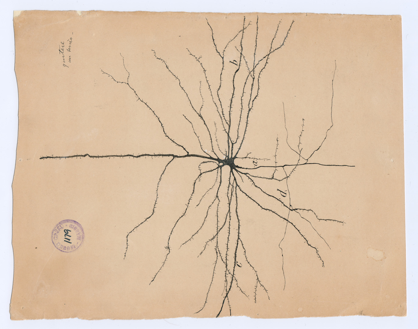
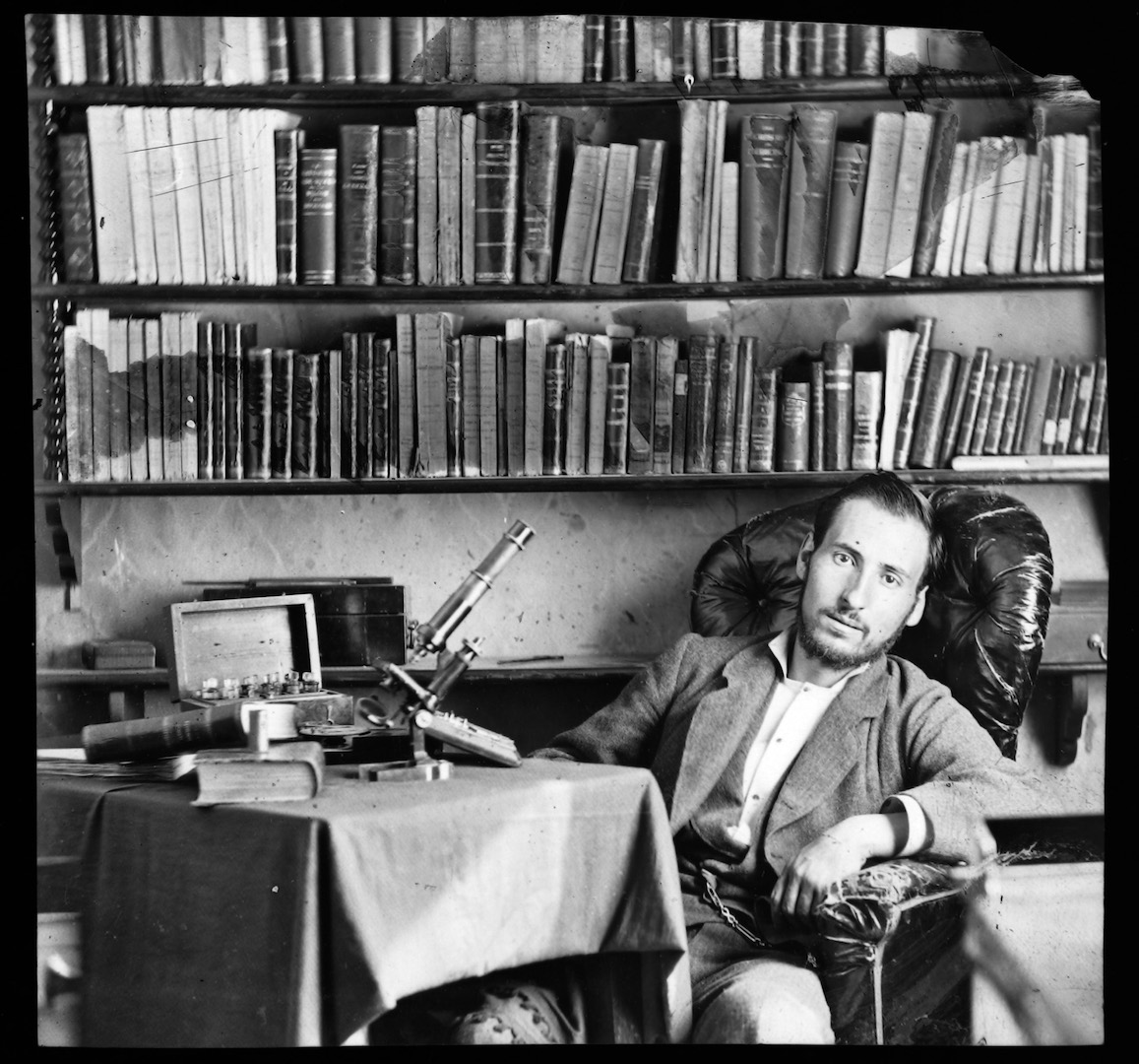
Santiago Ramón y Cajal was one of the first explorers to hack his way through the jungles of the mind. The Spanish Nobel laureate produced nearly 3,000 anatomical drawings of the microscopic brain—and these stark, mystifying illustrations are still shaping our understanding of that mysterious gray matter a century later. In fact, Cajal remains the most cited classical scientist in history.
Videos by VICE
Long hours crouched over a microscope, combined with Cajal’s self-described “irresistible mania for scribbling,” resulted in hundreds of detailed sketches of neurons, neuroglia, and all the other goop in our skulls. These illustrations are collected in a new book, The Beautiful Brain, published early this year. It traces the history of Cajal’s life and highlights the important discoveries he made. Flipping through these pages feels like breaking into Cajal’s lab.
Cajal was equal analyst and artisan, the author of more than 100 scientific articles, not to mention some pseudonymous science fiction under the name “Dr. Bacteria.” Fascinated by the photographic chemical process, Cajal was also a fervent photographer, even inventing some of his own color processing techniques.
But Cajal is known most of all for his intricate drawings of the brain. They illuminated our understanding of the nervous system and provided us with the neuron doctrine—arguably his greatest contribution to science—which declares the nervous system is assembled by individual cells.
Contrary to popular belief at the time, the nervous system is not a singular, self-contained, continuous network. It’s made up of discrete neurons, a cell type that transmits chemical and electrical impulses from one end to a neighboring neuron. A neuron receives these signals through its branch-like dendrites, sends it across a long projection called an axon and shoots it out the axon terminal, across a gap called the synaptic cleft, and into the dendrites of the next neuron. In other words, the mind is made up of individual neurons making up this chain reaction, as if they were “talking” to each other.
It wasn’t until the 1950s, around two decades after Cajal’s death, that he was proven correct. And he was quite poetic about his intentions: “To affirm that everything communicates with everything else is equivalent to declaring the absolute unsearchability of the organ of the soul,” Cajal once wrote.
Some of Cajal’s images are rough, as if erratically squiggled on scrap paper. But these drawings would have been printed in black and white, erasing pencil marks and pale correcting wax used to hide blemishes. Sometimes, Cajal exaggerated or darkened his images to emphasize his elaborate theories and even drew opposing ideas to demonstrate he was correct.
“That Cajal’s drawings remain living documents a century after they were created is at least partly owing to this vitality, which draws on fantasy and the imagination more than we might expect in a scientific project,” Lyndel King and Eric Himmel write in The Beautiful Brain.
Cajal had limited tools at his disposal, but could still see deeply. As mentioned, he predicted the path information flows between neuronal circuits. He looked at damaged pyramidal neurons and concluded that the brain cannot regenerate, unlike other nerves. He knew that astrocytes regulated the electrical responses of neurons, controlling their efficiency, which was only proven correct in the last 20 years.
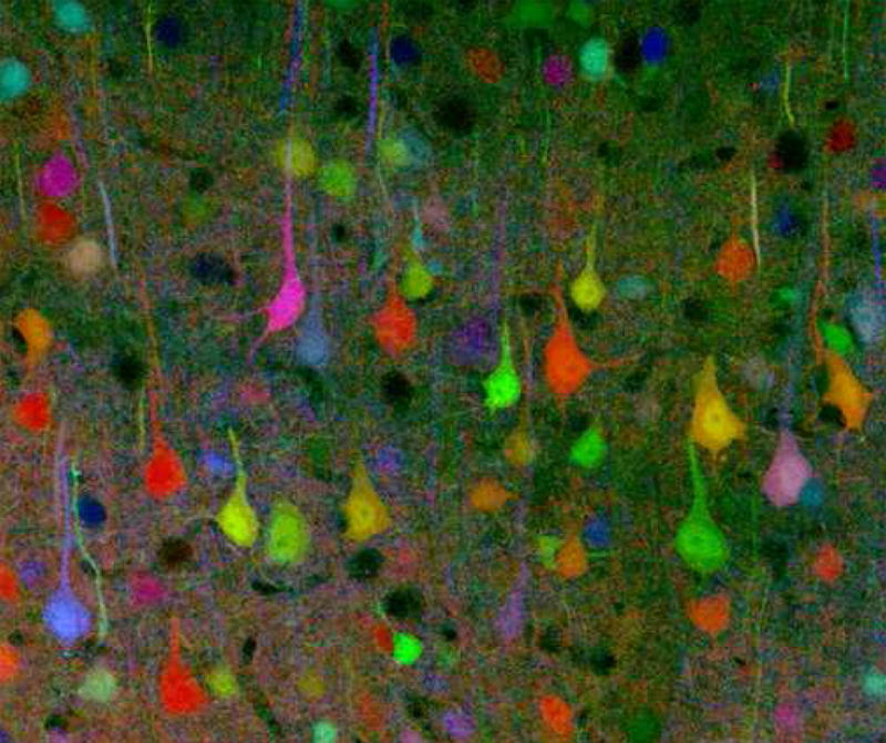
By improving Camillo Golgi’s silver-stain method, Cajal could darken and isolate nerve tissue under a microscope—otherwise, neurons are so densely woven, it’s almost impossible to pick them out from one another. (Golgi and Cajal shared a Nobel prize in 1906.) Today, we can use genetically-engineered rabies viruses that will dye some neurons red and others lime green to identify specific neural circuits.
This technique is more specific than the recently developed Brainbow, which manipulates the genes of the green fluorescent protein (GFP), found in a type of bioluminescent jellyfish. Modifying GFP’s allows researchers to splatter brain matter in living organisms with approximately 100 different fluorescent colors—literally a brain rainbow—making it easier to study neuronal relationships.
Indeed, we’ve come a long way from Angelo Mosso, who in the late 1870s became the first person to invent a neuroimaging technique by measuring the redistribution of cerebral blood flow. Today, one of the most advanced neuroimaging technique is positron emission tomography (PET), which creates 3D images by injecting radioactive tracers into the blood, then measures the resulting gamma rays.
The next major goal in neuroscience is a complete map of the brain, known as a connectome. As a group of researchers wrote in one Cell article, “for the most part, the precise relationships between the brain’s many cellular components are not known.” To turn turn images of brain into a mineable dataset, they cut a mouse brain into more than 10,000 slices less than 30 nanometers thick, which were then scanned by an electron microscope and reconstructed into 3D blobs that can show even synaptic vesicles.
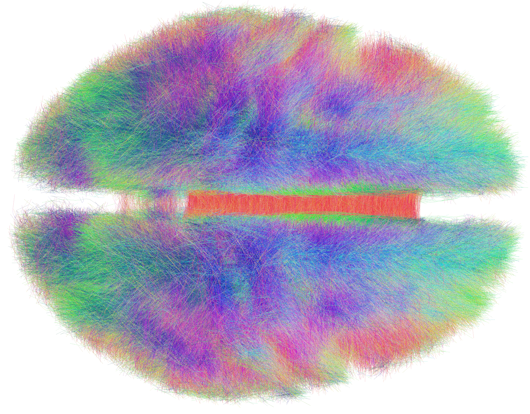
In order to build these computer models, they used VAST, a “computer-assisted manual space-filling segmentation and annotation program.” In other words, an artificial intelligence program. It’s a bit meta—using digital neural networks to map neural networks made of meat.
While this work is groundbreaking—literally the cutting edge of neuroimaging—it’s still tedious, hasn’t yet been applied to a human brain, and we still haven’t fully mapped these inner relationships. No one knows when we’ll get there, but China recently built a “brain-imaging factory,” last month the International Brain Lab opened, and groundbreaking research continues to come out a furious pace. In late October, for example, a team at Janelia Research Campus presented MouseLight NeuronBrowser, the “most extensive map of mouse brain wiring yet attempted.”
Today, we may understand the brain better than a century ago, but as Janet Dubinsky writes in The Beautiful Brain, “We do not know how the brain, three pounds of water, oil, small molecules, and protein can perform so many individual computations using so little energy. We do not fully understand how the brain creates the mind. In many ways, our twenty-first-century scientific questions and goals remain the same as Cajal’s: to ‘clarify the secret of mental life.’”
Get six of our favorite Motherboard stories every day by signing up for our newsletter .
More
From VICE
-
Photo: agung fatria / Getty Images -

Huge new game reveals at the 2025 Game Awards -

Photo: Yulia Reznikov / Getty Images -

WWE via Getty Images